Medisplint Medisplint

Why Choose Maxillofacial Plating Systems?

Time:2026-10-15 Author:Amelia
0%

Why Choose Maxillofacial Plating Systems?

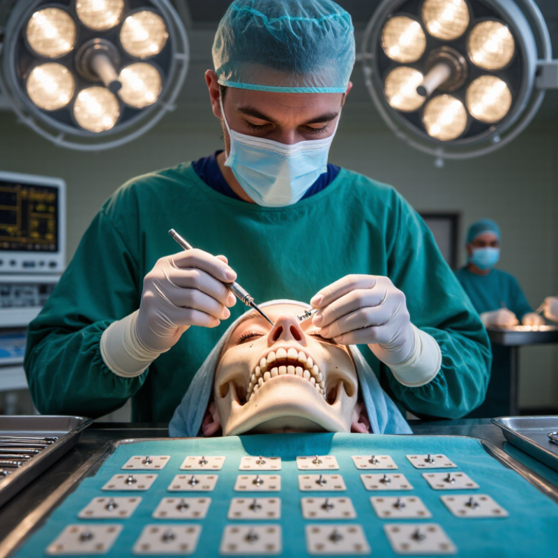
Maxillofacial Plating Systems support the precise stabilization of facial fractures and reconstructive procedures. They help surgeons align bone segments with controlled, predictable fixation. The right system can reduce movement during healing. It can also protect delicate anatomy near the orbit, nerves, teeth, and airway.

Dr. Edward Ellis III, a widely recognized oral and maxillofacial surgeon, has emphasized that facial fracture treatment must restore both form and function, rather than simply placing hardware. That principle remains important. A plate should follow the patient’s anatomy. Screws should provide dependable purchase without unnecessary irritation. Low-profile designs may improve comfort and reduce soft-tissue prominence. Clear labeling and compatible instruments can also support safer operating-room decisions.

Clinical experience still matters.

Surgeons assess fracture location, bone quality, occlusal relationships, imaging findings, and patient-specific risks before choosing a construct. Titanium systems are commonly valued for strength, corrosion resistance, and biocompatibility. Yet no implant solves every case. Complex comminution, infection risk, limited access, and unexpected anatomy can challenge even an experienced team. That is why reliable manufacturing, traceable quality control, and evidence-informed training deserve attention. A polished product description is not enough. Independent evaluation and long-term clinical feedback matter more.

The strongest reason to choose Maxillofacial Plating Systems is balance: stability without excess bulk, precision without unnecessary complexity, and innovation guided by patient outcomes. Good fixation is quiet. It supports healing while allowing the surgeon to focus on the person behind the scan.
Why Choose Maxillofacial Plating Systems?

What Are Maxillofacial Plating Systems and Their 1.5–2.4 mm Components?

Maxillofacial plating systems are small fixation platforms for repairing fractured facial bones. They usually combine contoured plates, locking or non-locking screws, drills, guides, and bending instruments. Common component thicknesses range from 1.5 to 2.4 mm. Thinner plates often support delicate orbital, nasal, and midface repairs. Thicker 2.0–2.4 mm plates generally provide greater stability for mandibular regions. The exact choice depends on bone thickness, fracture pattern, soft-tissue coverage, and load.

The need is substantial. The World Health Organization reported approximately 1.19 million road-traffic deaths worldwide in 2021, with another 20–50 million people suffering nonfatal injuries. Many of these injuries involve the face. Clinical guidance from the AO Foundation emphasizes three practical goals: restore anatomy, protect vital structures, and achieve stable fixation. A well-designed system can help surgeons place screws accurately through limited surgical exposure. It can also reduce unnecessary plate bending during reconstruction.

However, thickness alone should never determine selection. A 1.5 mm plate may be suitable in one region but inadequate under functional loading. A 2.4 mm plate may add strength, yet its profile can affect contouring and soft-tissue comfort. No system is perfect. Surgeons must still assess imaging, occlusion, bone quality, infection risk, and healing capacity. As reported in clinical literature, complications can include malocclusion, infection, hardware exposure, and nonunion. Careful planning matters more than impressive component numbers.

Why Choose Maxillofacial Plating Systems?

Maxillofacial plating systems use low-profile plates and fixation screws to stabilize facial bone segments. Common nominal screw diameters include 1.5 mm, 2.0 mm, and 2.4 mm. The chart compares their calculated circular cross-sectional areas using A = π(d/2)².

Note: These are nominal component diameters and geometric calculations, not clinical recommendations. Implant selection depends on anatomy, fracture pattern, bone quality, and the treating surgeon’s judgment.

Which Facial Fractures Require Fixation Across the Mandible, Midface, and Orbit?

Why Choose Maxillofacial Plating Systems?

Which Facial Fractures Require Fixation Across the Mandible, Midface, and Orbit?

Facial fracture fixation depends on stability, function, and displacement. Not every fracture needs a plate. A careful examination and thin-slice CT help guide the decision.

Mandibular fractures often require fixation when they cause malocclusion, segment movement, severe displacement, or multiple breaks. Fractures near the angle, body, or condylar region need particular assessment. Restoring the patient’s bite matters as much as restoring the bone contour. Small errors can become noticeable during chewing.

Midface fixation may be needed for displaced Le Fort fractures or unstable zygomaticomaxillary complex injuries. Signs include facial flattening, altered width, restricted mouth opening, or step-offs along the orbital rim. Stable, nondisplaced fractures may heal without surgery. The clinical threshold is not perfectly neat.

Orbital fractures require a functional review. Fixation is commonly considered with muscle entrapment, persistent double vision, globe displacement, or a defect that risks future sinking of the eye. Timing can change after swelling decreases. That detail is easy to overlook. Surgeons must also consider soft tissue injury, dental status, bone quality, and the patient’s general health.

A suitable plating system supports accurate reduction and stable healing across different facial regions. Plates should match the anatomy, while screws should provide secure purchase without entering vital structures. Yet no system compensates for poor exposure or incomplete reduction. Experience remains essential, and even experienced teams sometimes revise their plan after imaging or intraoperative findings.

Why Choose Maxillofacial Plating Systems? – Which Facial Fractures Require Fixation Across the Mandible, Midface, and Orbit?

Facial Region Typical Fracture Pattern When Fixation Is Commonly Considered Primary Treatment Objective Common Fixation Strategy Important Clinical Considerations
Mandible
Symphysis and Parasymphysis
Vertical or oblique fractures through the anterior mandible, including fractures near the incisor or canine region. Often requires fixation when displaced, unstable, open, associated with malocclusion, or unlikely to remain aligned with conservative care. Restore dental occlusion, mandibular continuity, alignment, and functional stability. One or more low-profile plates, load-sharing plates, or stronger load-bearing constructs depending on fracture pattern, bone quality, and stability. Assess tooth roots, mental nerves, soft-tissue injury, contamination, comminution, and the condition of the dentition before selecting screw positions.
Mandible
Body
Fractures extending between the canine and angle regions, frequently involving the tooth-bearing segment. Commonly considered for displaced, mobile, open, comminuted, or functionally unstable fractures. Re-establish occlusion and resist bending, torsional, and shear forces during healing. Miniplate fixation, two-plate configurations, or a load-bearing plate for severe instability, atrophic bone, or substantial comminution. Preserve the inferior alveolar nerve when possible; evaluate infection risk, teeth in the fracture line, bone stock, and postoperative occlusion.
Mandible
Angle
Fractures at the junction of the mandibular body and ramus, often influenced by the third molar region. Fixation is frequently used when there is displacement, malocclusion, instability, an open fracture, or inadequate control with closed treatment. Restore mandibular width, height, rotation, and occlusal relationship. A single superior border plate, angle plate, or more rigid construct selected according to fracture geometry and load requirements. Consider the third molar, infection risk, access limitations, masseteric forces, inferior alveolar nerve location, and soft-tissue coverage.
Mandible
Ramus and Condylar Region
Subcondylar, condylar neck, head, or ramus fractures; may be unilateral or bilateral. Open reduction and fixation may be considered for major displacement, loss of ramus height, persistent malocclusion, mechanical obstruction, or selected bilateral injuries. Preserve ramus height, mandibular movement, occlusion, and temporomandibular joint function. Small plates, specialized condylar plates, or alternative fixation methods based on access, fragment size, and surgeon preference. Evaluate facial nerve risk, joint involvement, intracapsular damage, mouth opening, occlusion, and the possibility of functional treatment without open surgery.
Midface
Le Fort I
Horizontal maxillary fracture above the tooth roots, separating the tooth-bearing maxilla from the midface. Usually requires fixation when displaced or mobile, particularly when occlusion and maxillary stability cannot be reliably maintained. Restore maxillary position, occlusion, facial height, and three-dimensional midface stability. Plates are commonly placed at accessible maxillary buttresses, often through intraoral approaches, after temporary occlusal stabilization. Confirm occlusion before definitive fixation; assess nasal, sinus, palatal, dental, and vascular injuries.
Midface
Le Fort II
Pyramidal midface fracture involving the nasal bridge, medial orbital region, infraorbital rims, and maxilla. Fixation is generally indicated when there is displacement, instability, malocclusion, orbital involvement, or loss of facial projection. Reconstruct the midface buttresses, orbital boundaries, nasal projection, and dental relationship. Multiple fixation points may be required at the nasofrontal, infraorbital, zygomaticomaxillary, and maxillary buttresses. Coordinate treatment of the orbit, nasal complex, lacrimal drainage system, airway, and occlusion; obtain thin-slice CT for planning.
Midface
Le Fort III
Craniofacial separation involving the zygomatic arches, lateral orbital walls, nasofrontal region, and entire midface. Typically requires rigid stabilization because of extensive displacement, craniofacial instability, and possible functional compromise. Re-establish craniofacial alignment, facial width and projection, orbital volume, and occlusion. Longer and multi-level fixation constructs are placed across the principal facial buttresses, often combined with craniofacial or orbital reconstruction. Prioritize airway, neurological assessment, cerebrospinal fluid leak evaluation, ocular injury, skull-base involvement, and associated brain trauma.
Zygomaticomaxillary Complex Fracture of the zygomatic body with possible involvement of the lateral orbital wall, orbital floor, infraorbital rim, and zygomatic arch. Fixation is commonly considered for malar flattening, displacement, trismus, orbital deformity, or loss of facial width and projection. Restore malar prominence, orbital shape, facial symmetry, and mandibular movement. One-, two-, or three-point fixation may be used at the frontozygomatic region, infraorbital rim, and zygomaticomaxillary buttress. Assess diplopia, enophthalmos, infraorbital nerve function, arch impingement, orbital volume, and the integrity of the zygomaticomaxillary buttress.
Orbit
Orbital Floor
Isolated or combined orbital floor fracture, including defects caused by blowout injury or extension from midface fractures. Repair is considered for persistent diplopia with evidence of entrapment, significant enophthalmos, hypoglobus, large defects, or progressive orbital volume change. Restore orbital volume and contour while releasing entrapped tissue and protecting ocular function. A contoured orbital implant or plate may be used to bridge and support the reconstructed orbital floor, depending on defect size and anatomy. Urgent ophthalmic assessment is required for vision loss, globe injury, retrobulbar hematoma, or muscle entrapment; imaging should guide implant positioning.
Orbit
Medial Wall
Fractures involving the thin medial orbital wall, sometimes extending into the ethmoid sinus and naso-orbito-ethmoid region. Repair may be considered for clinically significant enophthalmos, persistent diplopia, tissue entrapment, or a defect affecting orbital volume. Restore medial orbital contour and prevent abnormal globe position or soft-tissue tethering. Thin, anatomically shaped orbital reconstruction material may be used when direct support is needed; small stable defects may be observed. Evaluate the medial rectus, ethmoid sinus, optic nerve, lacrimal system, and nasal complex; avoid placing fixation near critical orbital structures.
Orbit
Medial Wall and Floor
Combined orbital wall fractures with increased orbital volume and possible involvement of the infraorbital rim. Often requires reconstruction when deformity, diplopia, enophthalmos, hypoglobus, or extensive wall loss is present. Reconstruct the three-dimensional orbital boundaries and restore pre-injury globe position. Patient-specific contouring or shaped orbital support may be combined with infraorbital rim fixation when the rim is unstable. Accurate restoration of the posterior ledge and orbital boundaries is essential; assess ocular motility and postoperative vision closely.
Naso-Orbito-Ethmoid Complex Central midface fractures involving the nasal bones, medial orbital walls, frontal process of the maxilla, and medial canthal tendon region. Fixation is generally required for displaced, unstable, or comminuted injuries, especially with telecanthus or loss of nasal support. Restore nasal projection, intercanthal distance, medial orbital contour, and airway structure. Small plates, screws, wires, or combined reconstruction methods may be used around stable bony segments and canthal tendon attachment areas. Assess the medial canthal tendon, lacrimal drainage system, nasal septum, frontal sinus, and cerebrospinal fluid leak risk.
Frontal Bone and
Anterior Frontal Sinus
Fractures of the frontal sinus anterior table, posterior table, or associated frontal bone and orbital roof. Fixation is considered for displaced contour deformity, instability, exposed fracture segments, or associated orbital and cranial injuries. Restore forehead contour and protect or reconstruct the anterior cranial and orbital boundaries. Low-profile plates or mesh may stabilize the anterior table or orbital roof after debridement and structural assessment. Posterior table injury, nasofrontal duct obstruction, cerebrospinal fluid leak, intracranial injury, and infection risk require specialist evaluation.
Alveolar and
Dentoalveolar Segment
Localized fracture of the tooth-bearing alveolar process, sometimes involving multiple teeth or the palate. Fixation is considered when the segment is mobile, displaced, maloccluded, or cannot be stabilized with less invasive methods. Restore occlusion, segment alignment, periodontal support, and tooth-bearing anatomy. Small plates, screws, or temporary splinting may be selected according to fragment size, tooth roots, and soft-tissue conditions. Protect tooth roots and developing teeth, assess pulp and periodontal injury, and maintain a clean environment for healing.

Fixation decisions depend on fracture displacement, occlusion, functional impairment, soft-tissue injury, bone quality, patient health, imaging findings, and the surgeon’s clinical judgment. Emergency eye, airway, neurological, and vascular problems should be assessed before definitive fracture fixation.

How Do ASTM F67 and F136 Titanium Grades Support Implant Performance?

Maxillofacial plating systems depend on more than plate shape or screw design. Titanium grade strongly influences strength, flexibility, corrosion resistance, and tissue compatibility. ASTM F67 covers commercially pure titanium, valued for its ductility and formability. It is highly formable. This can help surgeons contour plates around complex facial anatomy during reconstruction.

ASTM F136 specifies titanium alloy Ti-6Al-4V ELI. Its higher strength-to-weight ratio can support fixation where mechanical demands are greater. More strength helps. However, stronger does not always mean better. A plate that resists bending may become harder to contour accurately, increasing the risk of poor adaptation. Careful design and handling remain essential.

ASTM standards define important chemical and mechanical requirements, but they do not guarantee clinical success. Reliable assessment should include material certificates, lot traceability, surface quality, manufacturing controls, and sterilization validation. Traceability matters. Surgical technique, screw placement, bone quality, loading, and patient healing also affect implant performance. A practical selection process compares F67 and F136 against the anatomical site and expected forces. No material choice is perfect. That limitation deserves honest consideration, especially when laboratory strength data may not reflect every real clinical condition.

How Do Plate Profiles, Screw Diameters, and Hole Counts Affect Stability?

Why Choose Maxillofacial Plating Systems?

Plate profile, screw diameter, and hole count directly influence fixation stability. A low-profile plate can reduce palpability and soft-tissue irritation, especially along the zygomatic arch or mandibular border. However, thinner plates usually provide less bending resistance. The AO Foundation Surgery Reference (2024) emphasizes matching plate strength to fracture load, bone quality, and functional forces. A 2021 biomechanical study also reported greater bending resistance with 2.4 mm screws than with 2.0 mm screws. Larger screws can improve purchase, but they require adequate bone volume and careful drilling.

Screw diameter is only one part of the construct. Hole count affects flexibility, screw placement, and load distribution. More holes offer useful adjustment during surgery, yet each opening interrupts the plate’s solid cross-section. A 2022 systematic review in the Journal of Cranio-Maxillofacial Surgery found that stability depended strongly on plate position, fracture gap, and fixation length, not diameter alone. That finding is easy to overlook. Real patients rarely match laboratory models.

Tips: Choose the lowest profile that still supports the expected load. Use larger-diameter screws when bone thickness permits. Place screws on both sides of the fracture when anatomy allows. Avoid filling every hole automatically; preserve useful flexibility and respect vital structures. Surgical judgment still matters more than a specification sheet.

How Are ISO 10993 Testing and Clinical Outcomes Used to Compare Systems?

Why Choose Maxillofacial Plating Systems?

How Are ISO 10993 Testing and Clinical Outcomes Used to Compare Systems?

Choosing a maxillofacial plating system requires more than comparing screw sizes or catalog images. ISO 10993 testing offers a structured way to examine biological safety. Evaluation may consider cytotoxicity, sensitization, irritation, systemic toxicity, and material-related risks. Test selection should match the implant’s tissue contact and expected duration. That detail matters. A plate touching oral mucosa faces a different exposure pattern from one buried near bone. Manufacturing residues, surface treatments, and sterilization can also influence the biological assessment. However, a favorable test report is not a guarantee of clinical success. Interpretation needs traceable methods, current standards, and qualified review.

Clinical outcomes add the patient-facing evidence that laboratory testing cannot provide. Surgeons may compare fracture stability, union, infection rates, soft-tissue irritation, palpability, and unplanned removal. Patient anatomy and surgical technique strongly affect these results. A difficult mandibular angle can expose weaknesses that a controlled test misses. Follow-up duration matters too. Early comfort may hide later loosening, while delayed healing may reflect smoking, trauma, or poor bone quality. Reliable comparisons use defined endpoints, adequate follow-up, and transparent reporting of complications. Numbers alone can mislead. A low removal rate may reflect short observation, not superior performance. The most credible choice connects ISO 10993 evidence with real clinical records while acknowledging uncertainty. That discipline is demanding.

FAQS

Which mandibular fractures commonly need fixation?

Fixation is often considered for malocclusion, severe displacement, segment movement, or multiple fracture lines. The bite matters. A small alignment error may become obvious while chewing.

Can stable midface fractures heal without fixation?

Yes, stable and nondisplaced fractures may heal without surgery. Displaced injuries may need fixation when facial flattening, altered width, restricted opening, or orbital rim steps appear. The decision is not perfectly neat.

When should an orbital fracture be evaluated for fixation?

Evaluation is important with muscle entrapment, persistent double vision, globe displacement, or a large structural defect. A sinking eye may develop later. Swelling can change the timing of treatment.

What information helps surgeons plan facial fracture fixation?

Careful examination and thin-slice CT provide essential guidance. Surgeons also assess soft tissues, dental condition, bone quality, and general health. Imaging can change the plan.

How does plate profile affect fixation?

A low-profile plate may reduce palpability and soft-tissue irritation. This can matter near the mandibular border or cheekbone arch. Thinner plates usually resist bending less effectively.

How do screw diameter and hole count influence stability?

Larger screws may improve purchase when bone thickness allows safe placement. More holes provide adjustment options, but they also interrupt the plate’s solid cross-section. More is not always better.

How should screws be positioned around a fracture?

When anatomy permits, screws should support both sides of the fracture. They must avoid teeth, nerves, the orbit, and other vital structures. Do not fill every hole automatically.

What do biological testing and clinical outcomes reveal about a plating system?

Biological testing can assess cytotoxicity, irritation, sensitization, and systemic risks. Clinical records add evidence about union, infection, loosening, irritation, and removal. Neither source proves everything. Follow-up length can change the interpretation.

Conclusion

Maxillofacial Plating Systems are specialized implant assemblies designed to stabilize facial bones after fractures or reconstructive procedures. Their components commonly include low-profile plates and screws in the 1.5–2.4 mm range, selected according to the anatomy and load requirements of the treatment area. Fixation may be needed for fractures involving the mandible, midface, zygomatic region, nasal area, or orbit, with system design adapted to each site’s structural and functional demands.

Material selection is also important for implant performance. Titanium grades such as ASTM F67 and ASTM F136 are valued for their strength, corrosion resistance, and compatibility with biomedical applications. Plate profile, screw diameter, and hole count influence contouring, fixation options, load distribution, and surgical flexibility. ISO 10993-based biocompatibility testing, together with clinical outcomes such as stability, healing, and complication rates, helps clinicians and healthcare providers compare systems using safety, performance, and patient-focused considerations.

Amelia

Amelia

Amelia is a seasoned marketing professional with a wealth of expertise in our company’s core offerings. With an unwavering passion for driving growth and innovation, she plays a pivotal role in shaping our marketing strategies and enhancing brand visibility. A key aspect of her responsibilities......