Medisplint
Choosing the right Mandibular Reconstruction Plates can influence stability, healing, function, and long-term patient comfort. The decision requires more than comparing product prices or plate thickness. Surgeons must assess the defect’s location, bone quality, occlusion, soft-tissue condition, and reconstruction method. A plate supporting a short lateral defect may differ greatly from one spanning the entire mandible.
Clinical experience matters.
Modern systems often include locking screws, patient-specific designs, and several thickness options. However, advanced features do not automatically guarantee better outcomes. Peer-reviewed evidence, validated mechanical testing, and the manufacturer’s instructions should guide selection. Imaging quality is also essential. A precise three-dimensional scan can reveal thin cortical areas, nerve pathways, and limited screw purchase before surgery.
Small details matter.
The most reliable choice balances mechanical strength with anatomical fit. Excessive rigidity may complicate contouring or increase soft-tissue irritation. A thin plate may improve comfort but provide insufficient support in a high-load region. These trade-offs deserve discussion among maxillofacial surgeons, reconstructive specialists, biomedical engineers, and the patient when appropriate. No plate is perfect, and even experienced teams may revise their assumptions after reviewing new imaging or intraoperative findings.
This guide examines material properties, plate geometry, fixation methods, surgical workflow, and quality considerations. It also explains how regulatory documentation, sterilization requirements, and supplier support affect practical reliability. The goal is not to promote one universal system. It is to help qualified clinical teams make a careful, evidence-informed decision for each mandibular reconstruction case.
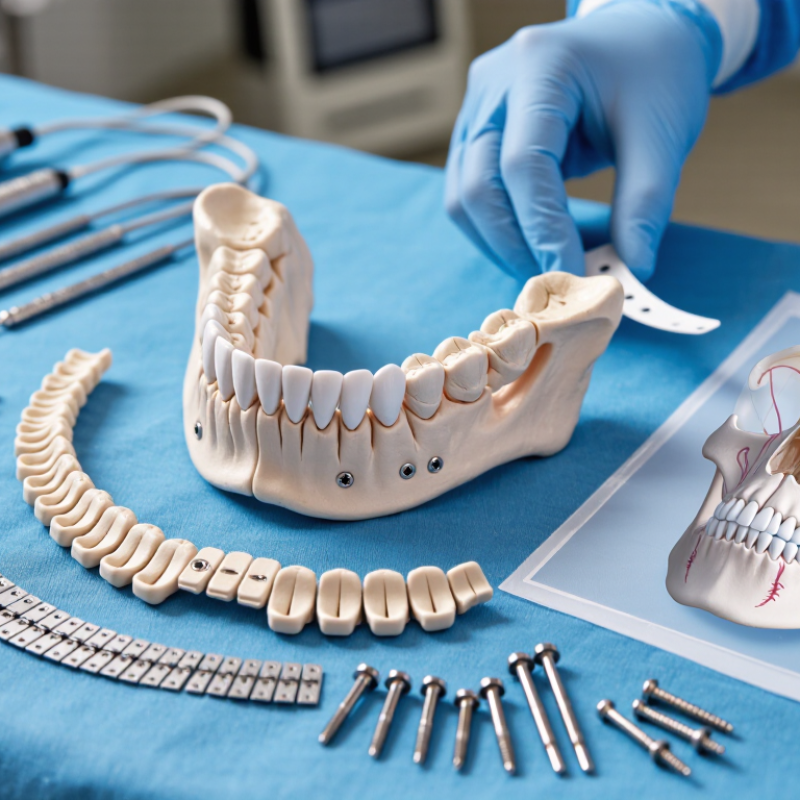
Mandibular reconstruction plates help restore jaw continuity after severe fractures, tumor removal, infection, or bone loss. They connect healthy bone segments and maintain the planned shape during healing. In many cases, the plate also supports a bone graft or vascularized tissue transfer. Its role is mechanical, but placement must respect nerves, teeth, vessels, and soft tissue.
Choosing a plate begins with the defect, not the product catalog. A surgeon reviews three-dimensional imaging, occlusion, bone quality, and the number of usable screw sites. A long segmental defect may need stronger load-bearing support. A smaller defect may allow a less bulky design. Low-profile plates can reduce soft-tissue irritation, although limited thickness does not always mean better comfort. Fit matters.
Screw direction and spacing deserve equal attention. Poor positioning can enter the tooth roots or the mandibular canal. Reconstruction should also preserve facial contour and allow practical oral care. In my experience, computer planning improves accuracy, yet it cannot replace direct assessment during surgery. A plate may fit the model but conflict with scarred tissue or unexpected bone loss. That is where judgment matters. Not always.
Patient factors can change the plan. Smoking, diabetes, previous radiation, nutrition, and infection may affect healing and fixation reliability. Follow-up imaging helps detect loosening, nonunion, or contour changes before symptoms become severe. Surgeons should match the plate’s strength and shape to the patient’s anatomy, healing capacity, and reconstructive objective. No selection rule is perfect. Reassessment remains essential.
Choosing the best mandibular reconstruction plate starts with patient anatomy, not catalog dimensions. The 2022 GLOBOCAN report estimated 389,846 new lip and oral cavity cancer cases worldwide. Many resections create asymmetric, irradiated, or contaminated surgical beds. CT imaging should show ramus height, cortical thickness, dental roots, and soft-tissue volume. Occlusion matters. Fit matters.
Defect size changes mechanical demand. A small lateral defect may need precise contouring and stable fixation near healthy bone. A long segmental defect creates greater bending forces, especially during chewing. A 2023 systematic review reported plate exposure rates commonly ranging from 10% to 25% across studies. Radiation, infection, poor soft-tissue coverage, and limited bone stock influenced outcomes. The figures vary. That variation deserves caution.
Reconstruction goals should guide the final choice. Restoring facial width may be the priority after tumor removal. Preserving occlusion, speech, swallowing, or future dental rehabilitation may matter more in another patient. Elderly patients with fragile bone require careful screw placement and gentle contouring. A younger patient may need a stronger pathway toward implant-supported function. I would not select a plate from defect length alone. A two-centimeter gap can behave unpredictably when the remaining bone is thin or poorly vascularized. The American Association of Oral and Maxillofacial Surgeons emphasizes individualized planning, yet real operating conditions can challenge even excellent imaging. Rechecking the occlusion and soft-tissue tension before final fixation remains a practical safeguard.
Choosing the best mandibular reconstruction plate requires more than selecting the strongest metal. Plate design controls how forces travel across the defect. Load-bearing plates should bridge the reconstructed segment when bone continuity is limited. Load-sharing designs depend more on graft or native bone support. Locking screw holes can preserve alignment when plate-to-bone contact is imperfect, but they may increase construct stiffness.
Material data helps explain the trade-off. ISO 5832-3 reports a minimum tensile strength of about 860 MPa for wrought titanium alloy used in implants. Stainless-steel implant standards report similarly high strength, often above 800 MPa. However, strength alone does not predict clinical performance. Titanium has a lower elastic modulus, near 110 GPa, than stainless steel, near 190 GPa. That difference may reduce stress shielding, although the plate’s thickness and cross-sectional shape often matter more.
Look closely at the contour. A plate with a deep profile resists bending better, while a thin plate may fit soft-tissue contours more comfortably. A 2023 systematic review in the International Journal of Oral and Maxillofacial Surgery linked plate exposure and fracture with factors such as radiation, poor soft-tissue coverage, and long unsupported spans. The evidence remains heterogeneous. It is not a perfect rule.
In practice, I would compare bending strength, fatigue resistance, screw locking behavior, and contour accuracy together. Bench testing should reflect repeated chewing, not only a single overload. ASTM F382 fatigue methods can guide testing, but laboratory results cannot fully reproduce infection, graft healing, or patient-specific forces. That limitation deserves attention.
Comparing representative ultimate tensile strength values for materials used or considered in mandibular reconstruction. Higher material strength can support thinner or more load-demanding designs, but clinical performance also depends on plate geometry, fixation, bone quality, and surgical loading.
Selecting the proper plate begins with anatomy, not appearance. The defect’s length, curvature, bone quality, and functional demands should guide the decision. Review recent CT images and dental occlusion before selecting a fixation plan. A plate must bridge the defect while preserving space for the planned graft. In clinical planning, virtual models can expose screw paths that conflict with tooth roots, vessels, or soft tissue. Measure twice. Small errors become difficult during surgery.
Tips: Map the defect in three dimensions. Check available bone for secure screw purchase. Keep screws away from roots and the inferior alveolar canal. Pre-contour the plate on a reliable model when possible. Prepare an alternative fixation plan. Another experienced review may reveal a missed weakness.
Surgical placement requires more than placing the plate against the mandible. Contour should follow the remaining bone without creating sharp pressure points. Confirm occlusion before final tightening, especially when reconstruction changes mandibular alignment. Use imaging and direct inspection to verify screw length, plate position, and graft space. Not every ideal plan survives exposure. Tissue swelling, limited access, or unexpected bone loss can change the operation. A flexible plan is safer than rigid confidence, although planning should remain precise and evidence-informed.
Fit matters early. A reconstruction plate should follow the planned mandibular contour without forcing the bone into position. Preoperative imaging and digital planning can reveal gaps, asymmetry, and weak screw sites before surgery. Surgeons should also assess occlusion, remaining bone quality, soft-tissue coverage, and the patient’s functional needs. A smooth fit may reduce bending during placement and support more predictable stability.
Stability depends on more than plate thickness. Screw distribution, bicortical purchase, segment length, and load sharing all influence fixation. The plate must resist chewing forces while the graft or bone heals. Yet excessive rigidity can create stress concerns in some situations. Careful intraoperative judgment remains essential. Small alignment errors can become visible when the patient bites, speaks, or opens the mouth.
Healing requires monitoring, not assumptions. Clinical reviews should examine wound condition, pain, infection signs, occlusion, and radiographic union. Nutrition, smoking status, previous radiation, and systemic disease may affect recovery. Long-term outcomes include speech, swallowing, facial symmetry, comfort, and the need for revision surgery. A plate that looks stable at six weeks may not deliver lasting function. That is an important limitation. Patient feedback should remain part of the assessment, because imaging cannot fully measure daily comfort or confidence.
Patient anatomy should guide selection, not catalog dimensions. Review CT images, bone quality, defect shape, and occlusion. Fit matters.
Examine ramus height, cortical thickness, dental roots, the inferior alveolar canal, and soft-tissue volume. These details influence plate position and screw safety.
Small lateral defects may need precise contouring and stable fixation near healthy bone. Long defects create greater bending forces during chewing. Size alone can mislead.
Occlusion affects mandibular alignment, speech, swallowing, and future dental rehabilitation. Check it before final tightening. Recheck it again after fixation.
Map screw paths in three dimensions. Keep screws away from tooth roots and the inferior alveolar canal. Verify screw length by imaging and direct inspection.
Pre-contouring on a reliable model can improve accuracy and shorten difficult adjustments. The contour should follow remaining bone smoothly. Avoid sharp pressure points.
Radiation, infection, poor soft-tissue coverage, and limited bone stock may increase plate exposure risk. Reported exposure rates vary widely. The figures need caution.
Facial width may matter most after tumor removal. Another patient may prioritize occlusion, swallowing, speech, or future implants. Fragile bone requires gentle contouring.
Prepare an alternative fixation plan before surgery. Swelling, limited access, or unexpected bone loss can alter the original plan. Flexible planning is safer.
No plan is perfect. Virtual models may reveal hidden conflicts, but operating conditions can still differ. An additional experienced review may expose missed weaknesses.
Choosing the best Mandibular Reconstruction Plates requires a careful balance between anatomy, surgical objectives, mechanical demands, and long-term healing. The process begins with evaluating the patient’s mandibular defect, bone quality, facial structure, occlusion, and the planned reconstruction method. These factors help determine the appropriate plate shape, size, thickness, hole configuration, and material. Plate designs should provide sufficient strength and stability while allowing accurate adaptation to the remaining bone and minimizing interference with surrounding tissues.
Successful selection also depends on detailed surgical planning and precise placement. The plate should support proper alignment, maintain functional stability, and accommodate any graft or flap used during reconstruction. After surgery, clinicians should assess fixation, fit, wound healing, bone union, comfort, and facial function over time. A well-matched plate can improve structural support and patient outcomes, but regular follow-up remains essential for identifying complications and confirming durable mandibular recovery.